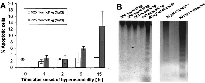
Figure 4.
Apoptosis cannot account for the increased DNA dsb during the early phase of hyperosmotic stress because its kinetics is slower than the induction of DNA dsb. (A) The percentage of apoptotic cells determined by neutral comet assay is low and not significantly different from isosmotic controls at all times when cells are exposed to 525 mosmol/kg HNa. Only after 15 h of exposure of cells to 725 mosmol/kg HNa does the number of apoptotic cells increase markedly. Data are means ± SEM (n = 3). (B) DNA ladder assay of mIMCD3 cells exposed to various degrees of HNa for 3 h. Equal amounts of whole genomic DNA were loaded in each well. Exposure of cells to 50 μg/ml etopside in serum-free medium served as a positive control for which the nucleosomal DNA ladder that is characteristic for apoptosis can be seen (last lane). DNA ladders are not present in genomic DNA of mIMCD3 cells exposed to HNa for 3 h, indicating that apoptosis is manifested at later times. Treatment with 25 μM LY294002 for 24 h did not induce apoptosis.

