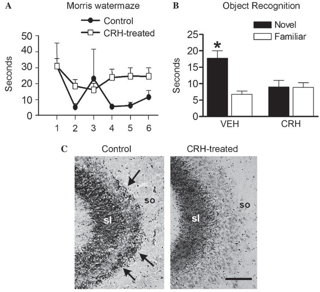
Fig. 6.
Administration of synthetic CRH into the brain recapitulates the structural and functional effects of early-life stress. (A) Adult rats that were treated with CRH early in life suffer from hippocampus-dependent memory dysfunction in the Morris watermaze. CRH-treated rats (white squares) take significantly longer to locate the hidden platform in the watermaze when compared to controls (black circles) [F(2,132) = 5.53, P< 0.01]. (B) Impaired memory is further evident by the performance of CRH-treated rats on the non-aversive, relatively stress-free object recognition test. Here, CRH-treated rats fail to distinguish the familiar object from the novel object, indicating impairment of recognition memory. (C) Sections of CA3A pyramidal cell regions from control and CRH-treated adult (12-month old), subjected to Timm’s stain for visualizing zinc-rich mossy fiber terminals. In CRH-treated rats, these terminals are abnormally abundant within the CA3 stratum oriens (so; arrows). sl, stratum lucidum. Scale bar = 50 µm. *P < 0.05. (Modified from [28] with permission.)

