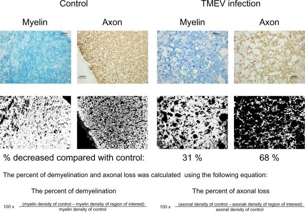
Fig. 2.
The percent of demyelination (myelin loss) and axonal loss during the chronic phase of TMEV infection. Normal axons were immunostained using an antibody cocktail against phosphorylated neurofilament, SMI 312, with diaminobenzidine (DAB) as the chromogen. Myelin was stained using Luxol fast blue. Densities of myelin sheaths and axons were compared in the white matter of the spinal cord between controls and TMEV-infected mice, using Image PloPlus. Sections of TMEV-infected spinal cord showed 31% myelin loss and 68% axonal loss, compared with control sections. This result supports the Inside-Out model of lesion development, whereby axonal damage heralds demyelination in TMEV infection. Scale bars = 20 μm.

