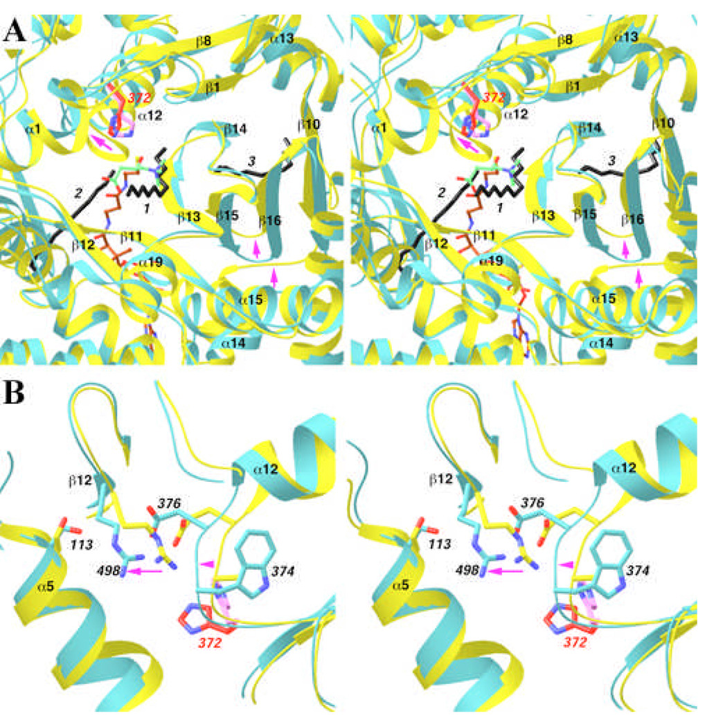
Fig. 5.
Structural differences between CPT-II and CrAT. (A). Overlap of the structure of CPT-II (in cyan) and CrAT (in yellow) near the binding site for the third aliphatic group. His372 in CPT-II is shown in red, and His343 in CrAT in magenta. Arrows in magenta indicate regions of conformational differences between the two structures. (B). Overlap of the structure of CPT-II (in cyan) and CrAT (in yellow) near the Ser113 residue. Produced with Ribbons [24].

