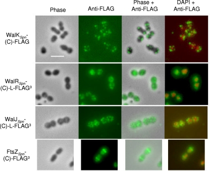
FIG. 2.
Localization of WalKSpn-(C)-FLAG, WalRSpn-(C)-L-FLAG3, and WalJSpn-(C)-L-FLAG3 by immunofluorescence microscopy (IMF) in exponentially growing unencapsulated strain D39. Representative phase images of fixed cells and IMF of the indicated FLAG-tagged proteins using anti-FLAG antibody are shown in the first and second columns, respectively. Carboxyl-terminal (C) fusion of the chromosomally expressed proteins to the linker (L) and FLAG epitope tags (Table 1) did not cause any observable changes in cell growth or morphology compared to parent strains (first column; data not shown). IMF of cells lacking a FLAG-tag fusion protein did not show any labeling (data not shown). The third and fourth columns show overlays of the first two columns or overlays of the second column and images stained with DAPI to locate nucleoids, respectively. For comparison and to validate the IMF methods, the bottom row shows localization of FtsZSpn-(C)-FLAG3 at the equators of exponentially growing cells of R6, which is an unencapsulated laboratory strain originally derived from strain D39. At least three biological replicates were performed for each strain, and multiple fields of cells were observed and analyzed for each replicate. See Materials and Methods for the IMF staining procedure used and the text for additional details.

