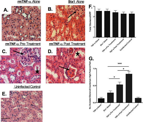
FIG. 4.
TNF-α pre- and posttreatment alter Shiga toxin 1-mediated renal histopathology. Mice were treated with rmTNF-α alone (A), 9 μg/kg Stx1 alone (B), pretreated with rmTNF-α 24 h before toxin (C), posttreated with rmTNF-α 24 h after toxin challenge (D), or treated with saline (E). Kidney halves were harvested 48 h after toxin administration and processed for H&E analysis. Slides were blinded, examined, and quantified by a veterinary pathologist. Arrows in panels B, C, and D point to glomeruli; stars in C and D denote renal tubule damage. The magnification of all images is ×40 (bar = 500 μm). Glomerular quantification was done blinded by counting total glomeruli (F) versus hyperemic glomeruli (G) per high-power field (10 fields were counted per slide). The experiment was repeated twice with a total of six mice. Statistical significance was assessed by one-way ANOVA and Tukey's multiple comparison post hoc test. An asterisk denotes significance at P < 0.05, and three asterisks denotes significance at P < 0.001.

