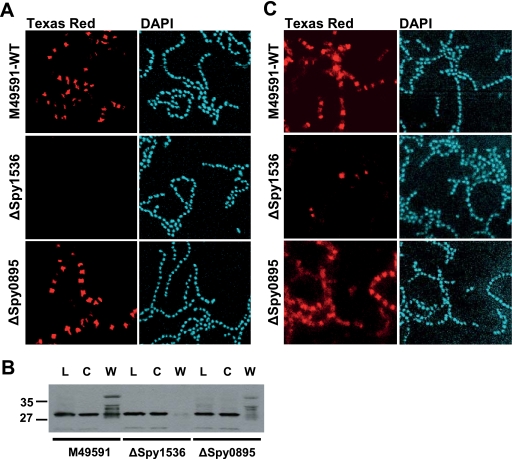
FIG. 6.
Localization of M protein and Spy0269 on the surfaces of wild-type (WT), ΔSpy1536, and ΔSpy0895 GAS cells. (A) S. pyogenes cells of an overnight culture of M49591 and ΔSpy1536 and ΔSpy0895 gene deletion mutants were subjected to immunofluorescence analysis with M23-specific polyclonal mouse antibodies, Texas Red dye-conjugated goat anti-mouse antibodies, and DAPI, following microscopic visualization. (B) Western blot analysis of cell fractions of wild-type M49591 and ΔSpy1536 and ΔSpy0895 cells to detect M protein, total lysate (lanes L), the cytoplasmic fraction (lanes C), and the cell wall fraction (lanes W) with M1 protein-specific polyclonal mouse antibodies. For all fractions, an amount of ∼30 μg total protein as measured by the BCA assay was loaded. Numbers at the left indicate molecular weights (in thousands). (C) Immunofluorescence analysis of WT M49591 and ΔSpy1536 and ΔSpy0895 gene deletion mutants with Spy0269-specific polyclonal mouse antibodies, Texas Red dye-conjugated goat anti-mouse antibodies, and DAPI.

