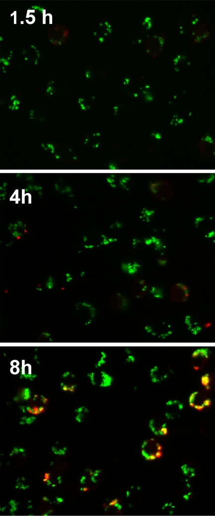
FIG. 1.
Determination of extracellular versus intracellular Y. pestis associated with macrophages. J774A.1 cells were infected with KIM5/pGFP at an MOI of 50 and fixed with 2.5% paraformaldehyde at the indicated time points. GFP expression was induced 1 h prior to fixation. The fixed but nonpermeabilized cells were treated with anti-Yersinia antiserum SB349 (48) to specifically label extracellular yersiniae. The slides were examined by epifluorescence microscopy to identify the macrophage-associated intracellular bacteria (green due to GFP only) and extracellular bacteria (red and green overlay). Three independent experiments were performed, and representative pictures are shown.

