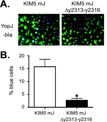
FIG. 7.
Determination of Yop translocation using bla assay. BMMs were infected with Y. pestis strains (KIM5 mJ or KIM5 mJ Δy2313-y2316) expressing YopJ-bla (Table 1) at an MOI of 10. The CCF2-AM dye was added at 4 h postinfection. The cells were incubated for an additional hour, and images were taken using fluorescent microscopy. Bacteria were grown at 37°C for 2 h in the presence of 2.5 mM calcium prior to macrophage infection. (A) Representative pictures of the infected BMMs. Blue indicates the translocation of the Yop-bla fusion protein into the BMMs. (B) Percent blue cells. The results are the averages of three independent experiments, in which cells were counted in three independent fields. Error bars represent standard errors of the mean (SEMs). The asterisk indicates a significant difference compared to the result for the wild type (P < 0.05), as determined by Student's t test.

