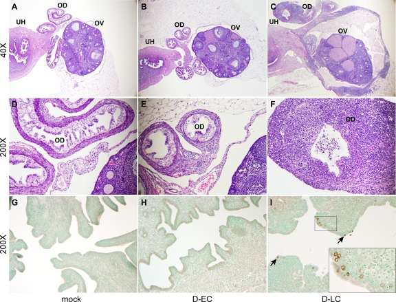
FIG. 3.
Histopathological evaluation of oviduct and ovarian tissue from C. trachomatis-infected mice. (A to F) Micrographs are of representative examples of H&E-stained tissues evaluated at 42 days p.i. from mock-, D-EC-, and D-LC-infected mice at ×40 (A to C) and ×200 (D to F) magnifications. The indicated tissue types are uterine horns (UH), oviducts (OD), and ovaries (OV). The oviduct and ovarian tissues of mock-infected (A and D) and D-EC-infected (B and E) mice are of normal thickness and contain only minimal aggregates of perivascular lymphocytes. In contrast, the walls of the oviducts and ovarian bursa of D-LC-infected mice (C and F) are severely thickened by lymphocyte and plasma cell infiltrates. Proteinaceous fluid in the lumen of the oviducts and ovarian bursa primarily consisted of polymorphonuclear leukocytes and was observed in both mock-infected and infected mice. (G to I) Uterine horn tissues from mock-, D-EC-, and D-LC-infected mice stained at 42 days p.i. with anti-MOMP-specific monoclonal antibody L2I-5 (magnification, ×200). Chlamydiae were not detected in the tissues of mock-infected (G) or D-EC-infected (H) mice but were readily found in the tissues of D-LC-infected mice (I). Inclusions (arrows) were localized to the epithelial cells. (I, inset) Enlarged image of immunostained inclusions.

