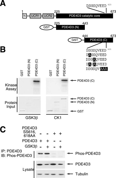
FIG. 6.
CK1 and GSK3β phosphorylate PDE4D phosphodegron. (A) Schematic illustration of PDE4D3 and PDE4D3 GST fusion protein. The location of the DSGSQVEED phosphodegron in PDE4D3 are also shown. Replacements of critical amino acid residues in the DSGSQVEED phosphodegron of PDE4D3 with Ala are highlighted. (B) GST recombinant proteins encompassing either the NH2-terminal or the COOH-terminal portion of the catalytic core of PDE4D3 were subjected to in vitro protein kinase assays in the presence of recombinant GSK3β or CK1 and [γ-32P]ATP. Autoradiography and Coomassie blue staining of recombinant PDE4D3 are shown. (C) COS7 cells transiently transfected with wild-type or Ala616,618 mutant PDE4D3. The effect of GSK3β was also examined. Phosphorylation of Ser616 of PDE4D3 was assessed by phospho-PDE4D antibody (Phos-PDE4D) in immunoblot analysis. Expression of PDE4D3 and tubulin is also shown.

