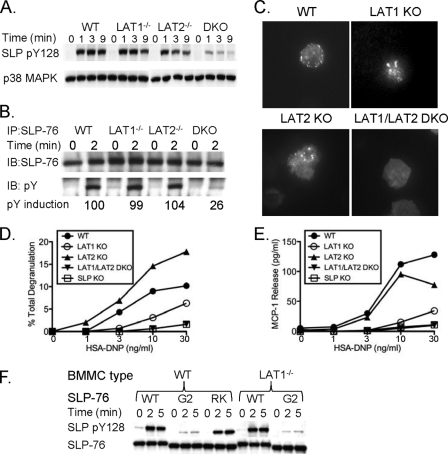
FIG. 3.
LAT2 can support SLP-76 phosphorylation and membrane recruitment in the absence of LAT1. (A and B) WT, LAT1−/−, LAT2−/−, and LAT1−/− LAT2−/− (double knockout [DKO]) BMMCs were preincubated with anti-DNP IgE (1 μg/ml) and stimulated with HSA-DNP (30 ng/ml) for the indicated time. Cell lysates were analyzed for phosphorylated Y128 of SLP-76 (pY128 SLP) with a pY128-specific antibody and total SLP-76 (A) or immunoprecipitated (IP) with anti-SLP-76 antibody and immunoblotted (IB) for phosphotyrosine (pY) and total SLP-76 (B). (C) Anti-DNP IgE-presensitized GFP-SLP-76-transduced WT, LAT1−/−, LAT2−/−, and LAT1−/− LAT2−/− BMMCs were deposited onto anti-IgE antibody-coated coverslips, and GFP-SLP-76 clustering at the plasma membrane was monitored by TIRF microscopy. Twenty cells were visualized per treatment, and images are representative of four independent experiments. (D and E) WT, LAT1−/−, LAT2−/−, LAT1−/− LAT2−/−, and SLP-76−/− BMMCs were preincubated with anti-DNP IgE and stimulated with various concentrations of HSA-DNP. Cell-free culture supernatants were analyzed for degranulation (D) and MCP-1 production (E). One representative of at least three independent experiments is shown for each panel. (F) WT, SLP-76.G2 (G2) mutant, or SLP-76.RK (RK) mutant GFP-SLP-76-transduced WT or LAT1−/− BMMCs were preincubated with anti-DNP IgE (1 μg/ml) and stimulated with HSA-DNP (30 ng/ml) for the indicated time. Cell lysates were analyzed for phosphorylated Y128 of SLP-76 (pY128 SLP) and total SLP-76. One representative of two independent experiments is shown.

