FIG. 4.
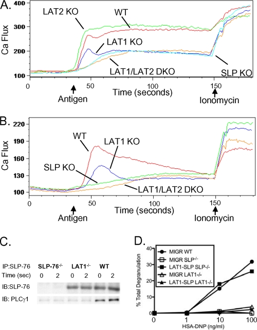
Cooperation of SLP-76 and LAT1 is important for FcɛRI-mediated signaling and function. (A) WT, LAT1−/−, LAT1−/− LAT2−/−, and SLP-76−/− BMMCs were preincubated with anti-DNP IgE (1 μg/ml) and monitored for elevations in intracellular Ca2+ by flow cytometry after stimulation in Tyrode's buffer. (B) WT, LAT1−/−, and SLP-76−/− BMMCs were preincubated with anti-DNP IgE (1 μg/ml) and monitored for elevations in intracellular Ca2+ by flow cytometry after stimulation in PBS containing EGTA (10 mM). The arrows indicate the time when the stimulus (HSA-DNP or ionomycin) was added to the mast cells. (C) WT, LAT1−/−, and SLP-76−/− BMMCs were preincubated with anti-DNP IgE and stimulated with HSA-DNP (30 ng/ml) for the indicated time. Cell lysates were immunoprecipitated with anti-SLP-76 antibody and blotted for total SLP-76 and PLCγ1. (D) WT, SLP-76−/−, or LAT1−/− BMMCs were retrovirally transduced with vector alone (MIGR) or full-length SLP-76 fused to the extracellular domain of LAT1 (LAT1-SLP), preincubated with anti-DNP IgE, and stimulated with various concentrations of HSA-DNP. Cell-free culture supernatants were analyzed for degranulation. Results are representative of at least two independent experiments.

