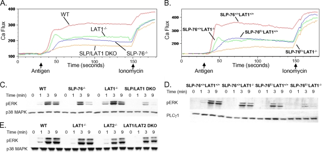
FIG. 5.
SLP-76 and LAT1 contribute independently to Ca2+ flux and ERK phosphorylation. (A and B) WT, LAT1−/−, SLP-76−/−, and SLP-76−/− LAT1−/− BMMCs (A) or YFP-positive SLP-76+/+ LAT1+/+, SLP-76+/+ LAT1−/−, SLP-76F/− LAT1+/+, and SLP-76F/− LAT1−/− BMMCs (B) were preincubated with anti-DNP IgE (1 μg/ml) and monitored for elevations in intracellular Ca2+ by flow cytometry. The arrows indicate the time when the stimulus (HSA-DNP or ionomycin) was added to the mast cells. (C and D) WT, LAT1−/−, SLP-76−/−, and SLP-76−/− LAT1−/− BMMCs (C) or YFP-positive SLP-76+/+ LAT1+/+, SLP-76+/+ LAT1−/−, SLP-76F/− LAT1+/+, and SLP-76F/− LAT1−/− BMMCs (D) were preincubated with anti-DNP IgE and stimulated with HSA-DNP (30 ng/ml) for the indicated time. Cell lysates were analyzed for phosphorylated ERK (pERK), total p38 MAPK, or total PLCγ1 by Western blotting. (E) WT, LAT1−/−, LAT2−/−, and LAT2/LAT1 DKO BMMCs were preincubated with anti-DNP IgE (1 μg/ml) and stimulated with HSA-DNP (30 ng/ml) for the indicated time. Cell lysates were analyzed for phosphorylated ERK and total p38 MAPK. Total p38 MAPK or PLCγ1 served as loading controls. Results are representative of at least two independent experiments.

