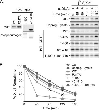
FIG. 4.
XCdt2 promotes the turnover of Xic1 in the egg extract. (A, right) Xic1 degradation assay. 35S-Xic1 was incubated in the HSS supplemented with XB− buffer, unprogrammed reticulocyte lysate (Unprog. Lysate), XCdt2WT (WT), XCdt2R247A (R247A), XCdt21-400 (1-400), XCdt2401-710 (401-710), or both XCdt21-400 and XCdt2401-710 with (+) or without (−) single-stranded DNA (ssDNA), and samples were analyzed at time points between 0 and 180 min as indicated. (Left) Input amounts of unlabeled in vitro-translated XCdt2 proteins added were quantitated by Western blotting (WB) using anti-Cdt2 antibody. 35S-labeled Cdt2 (1-400) was also quantitated by phosphorimager analysis since it is not recognized by the anti-Cdt2 antibody which was generated against the C-terminal fragment of Cdt2. (B) Quantitation of Xic1 turnover. The mean percentage of Xic1 remaining from two or three independent experiments as described in the legend to panel A is shown, where the 0 h time point was normalized to 100% of Xic1 remaining for each sample. SEMs are shown as error bars.

