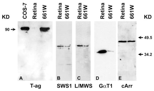
Figure 1.
Comparison of pattern of expression of cone-specific antigens in 661W cells to that of normal retina. Aliquots of 20 μg protein in extracts from retinas and 661W cells were electrophoresed, transferred to PVDF membrane, and probed with antibodies against SV40 T-ag (T-ag, A), blue cone opsin (SWS1, B), red-green cone opsin (L/MWS, C), transducin (GαT1, D), and cone-rod arrestin (cArr, E). Left: molecular mass of T-ag; right: migration of two protein molecular mass markers.

