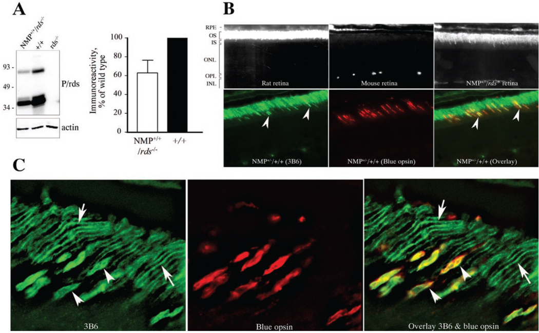
FIGURE 1.
NMP protein levels and OS localization. (A) Immunoblot analysis of retinal extracts from NMP+/+/rds−/−, wild-type (+/+), and rds−/− mice. Note the expected absence of P/rds immunoreactivity in the rds−/− retinal extract. Densitometric measurements of four independent blots are shown and were used to assess levels of NMP protein at 60% of endogenous P/rds. (B) C-terminal P431Q modification allowed immunofluorescent recognition of the NMP protein by 3B6 antibody. Top: ability of mAb 3B6 to recognize rat P/rds and NMP, but not endogenous mouse P/rds. Positively labeled retinal sections (rat and NMP) show mAb 3B6-staining in OS. Bottom: NMP retinal sections stained with both mAb 3B6 and anti-blue cone opsin antibodies, demonstrating colocalization of immunofluorescent signals. Arrowheads: localization to cone OS. (C) At higher magnification, single staining with mAb 3B6 and double staining with mAb 3B6 and anti-blue cone opsin antibodies further confirmed appropriate localization of the NMP protein to the OS disc rims of both rods (arrows) and cones (arrowheads).

