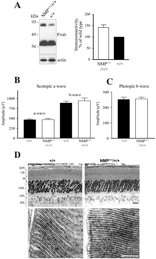
FIGURE 5.
Effect of P/rds overexpression on photoreceptor function and structure. (A) Blot analysis demonstrated the overexpression of P/rds in NMP+/+/+/+ retinas relative to wild type. Densitometry scanning of blots revealed a level of P/rds equivalent to approximately 150% of wild type. (B) Scotopic and (C) photopic ERG analyses showed no deleterious effects of P/rds overexpression on photoreceptor function up to 7 months of age, the latest time assessed. ERG wave amplitudes represent an average of 12 to 16 eyes for each genotype. (D) Histologic and ultrastructural appearances, including outer nuclear layer count (9 – 10 rows), are comparable in retinas from 4-month-old NMP+/+/+/+ and wild-type mice, indicating the lack of negatives effects associated with P/rds overexpression in the mouse retina. Scale bare: (D, top) 20 µm; (D, bottom) 0.2 µm.

