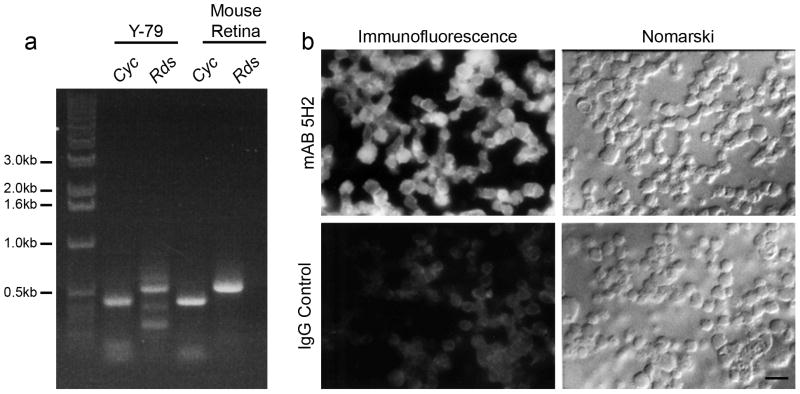
Figure 2. Expression of the mouse Rds gene in Y-79 cell lines.
a. Expression of the Rds gene detected by RT-PCR. Primers used for each PCR reaction are indicated on top of each lane (Rds, Cyc). The size of the products from Rds and cyclophilin gene (control) are also labeled. b. Expression of the Rds gene detected by immunocytochemistry using mAB 5H2 against RDS. Immunostaining was observed in the majority of the Y-79 cells (top left) while no staining was seen in IgG stained control cells. Scale bar 25 μm.

