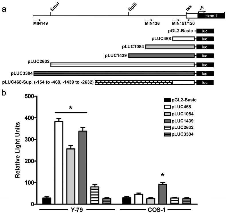
Figure 3. Promoter activity of the 5′ flanking region of the mouse Rds gene.
a. Schematic showing the construction of the luciferase reporter constructs. b-c. Luciferase activity (normalized to β-gal) after co-transfection of 15 μg of the indicated luciferase construct (containing different Rds promoter fragments) and 10 μg of pCH110 (used as an internal control) into Y-79 and COS-1 cells. b. Three of the constructs drove significant expression after transfection into human Y-79 retinoblastoma cells (left) while little expression was detected after transfection into COS-1 cells (right). The data shown is an average of three independent experiments, * p<0.01.

