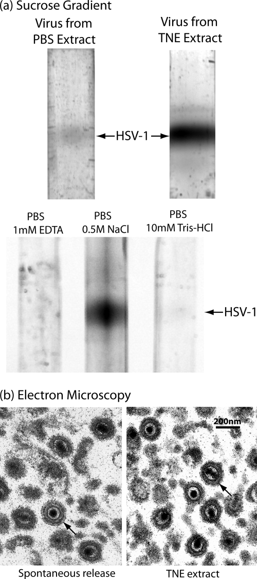
FIG. 1.
Characterization of HSV-1 harvested from the infected cell surface by treatment with TNE. (a) Sucrose density gradient ultracentrifugation and (b) electron microscopy are shown. The top two gradients in panel a compare the amounts of virus released by treatment of cells infected for 18 h with TNE (right gradient) and PBS (left). Note that substantially more virus was released with TNE. The lower three gradients in panel a compare the amounts of virus released with PBS containing the individual components of TNE, 1 mM EDTA, 0.5 M NaCl, and 10 mM Tris-HCl, pH 7.4. Note that the amount of virus released was greatest with PBS containing 0.5 M NaCl. Micrographs in panel b show HSV-1 spontaneously released from Vero cells infected for 18 h (left) and HSV-1 released from similar cells by treatment with TNE. Note that in both cases the tegument has the uniform arrangement characteristic of early-release virus (27). Arrows indicate representative virions in both cases.

