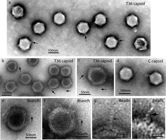
FIG. 3.
Electron microscopy of T36 capsids (a to c and e to h) and nuclear C capsids (d). All specimens were prepared by negative staining. Heavily stained images are shown in panel a, while other specimens are more lightly stained. Note that in all cases, T36 capsids had projections not found in C capsids. Projections appeared as tufts after heavy staining (a) and strands when staining was lighter (b, c, and e to h). Some strands were found to branch, as shown in panels e and f. Substructure was observed in strands, suggesting a coiled or beaded composition (g and h).

