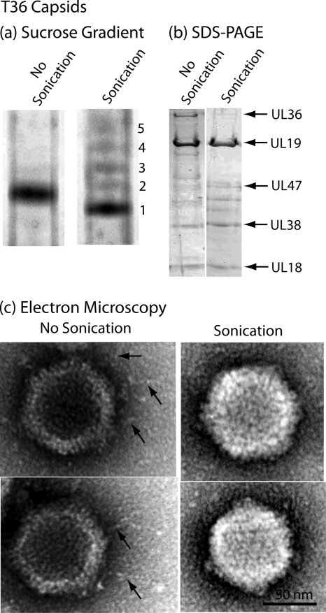
FIG. 5.
Characterization of sonicated T36 capsids by sucrose density gradient centrifugation (a), SDS-polyacrylamide gel electrophoresis (b), and electron microscopy (c). For the procedures used to produce T36 capsids and to expose them to brief sonication, see Materials and Methods. Note that sucrose density gradient analysis demonstrated that sonication resulted in the formation of a predominant band of capsids (band 1) that migrated slightly more rapidly than control, unsonicated T36 capsids. Minor bands of more slowly migrating capsids are suggested to have lost DNA as a result of sonication. SDS-polyacrylamide gel and electron microscopic analyses demonstrated that sonication caused loss of UL36 and UL37 and also of the fibers projecting from the T36 capsid surface. The findings are interpreted to support the view that the fibers are composed of UL36 and/or UL37.

