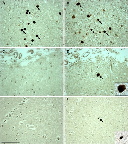
FIG. 2.
In situ hybridization (ISH) detection of JCV-positive cells in the brain samples of HIV-positive PML, HIV-positive, and HIV-negative individuals compared to the immunohistochemical staining detection of JCV VP1 proteins in the same patients. Positive VP1 protein staining in JCV-infected cells (arrows) in an HIV-positive PML individual (A) corresponds to positive ISH for JCV DNA (arrows) in the same individual (B). (C) JCV VP1 protein is not detected in the brain of an HIV-positive individual. (D) However, ISH detected rare cells harboring JCV DNA (arrow) in this patient. (E) JCV VP1 protein is not detected in an HIV-negative individual. (F) However, ISH showed rare JCV DNA-positive cells (arrow). The images are magnified 40-fold, and the insets are magnified 100-fold. Scale bar = 100 μm.

