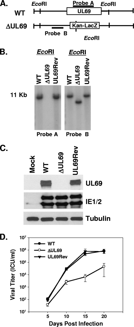
FIG. 1.
Generation and characterization of the UL69 deletion and UL69 revertant viruses. (A) Schematic representation of WT HCMV BAC and ADΔUL69 BAC depicting insertion of the Kan-LacZ cassette and the EcoRI restriction sites. (B) Southern blot analysis of WT pADCREGFP, pADΔUL69 (ΔUL69), and pADUL69Rev (UL69Rev) BACs digested with EcoRI restriction enzyme and probed for either UL69 (probe A) or the UL69 left flanking region (probe B). (C) Western blot analysis of UL69 expression at 72 h postinfection in HFF cells that were either mock infected or infected with WT, ADΔUL69 (ΔUL69), or ADUL69Rev (UL69Rev) virus. Lysates were also assayed for the abundances of IE1 and IE2. Anti-tubulin (α-tubulin) abundance was included as a loading control. (D) HFF cells were infected (0.01 PFU/cell) with either WT ADCREGFP, ADΔUL69, or ADUL69Rev virus. Cultures were harvested at the indicated times postinfection, and infectious virus was quantified by infectious-center assay. Results represent three independent experiments. ICU, infectious-center units.

