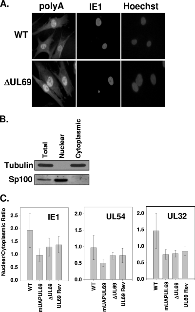
FIG. 4.
Expression of UL69 does not affect shuttling of RNA during HCMV infection. (A) HFF cells were infected at an MOI of 0.5 PFU/cell with either WT or ADΔUL69 (ΔUL69) virus. Seventy-two hours postinfection, cells were fixed and assayed for mRNA localization within the cells by in situ hybridization, using a biotinylated oligo(dT) probe, and were visualized by fluorescence staining with streptavidin-Alexa Fluor 546-conjugated antibody, followed by immunofluorescence staining for the HCMV IE1 protein. (B) HFF cells were infected with either WT, ADmUAPUL69 (mUAPUL69), ADΔUL69 (ΔUL69), or ADUL69Rev (UL69Rev) virus at an MOI of 1 PFU/cell. Seventy-two hours postinfection, cells were harvested, and nuclear and cytoplasmic fractions were isolated using a PARIS kit (Ambion) according to the manufacturer's instructions. Total, nuclear, and cytoplasmic proteins were separated via SDS-PAGE, transferred to nitrocellulose membranes, and subjected to Western blot analysis using antibodies directed against either the cytoplasmic protein α-tubulin or the nuclear protein Sp100. (C) Quantitative PCR was performed on cDNA generated from both the nuclear and cytoplasmic fractions of cells infected with the indicated viruses. Primers and probes specific to an immediate early (IE1), an early (UL54), and a late (UL32) gene were used to amplify and quantify the amount of each transcript present in each fraction. No statistical difference (all P values were >0.2) in the ratios was observed for any of the viral infections.

