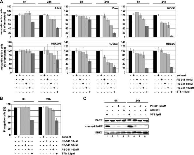
FIG. 1.
A 50 nM concentration of PS-341 is not cytotoxic or proapoptotic in cells for the indicated exposure times. A549 (A and B) and Vero, MDCK II, and HEK293 cells and HUVEC and HBEpC (all shown only in panel A) were treated with different concentrations (100 nM, 50 nM, and 10 nM) of PS-341 for the indicated times or treated with solvent or left untreated. Cells treated with 1.5 μM staurosporine (STS) were used as positive controls for metabolically inactive cells/dead cells. (A) An MTT assay was performed, and metabolic activity of cells was calculated as the percentage of the untreated control. Arrow bars represent standard deviations from four independent experiments. (B) PI staining was performed to measure membrane integrity of cells by fluorescence-activated cell sorter analysis. The diagram shows gated cells which were not stained by PI and therefore had no destruction of the cell membrane. Arrow bars represent standard deviations of three independent experiments. (C) A549 cells were treated with 50 nM PS-341 or solvent or left untreated for the indicated times. Afterwards cells were lysed and Western blot analysis was performed to detect apoptotic cells by cleavage of PARP. As a positive control cells were treated with 1 μM staurosporine. Equal amounts of protein loading were assayed by using the cellular protein ERK2. Shown is one representative blot out of three independent experiments.

