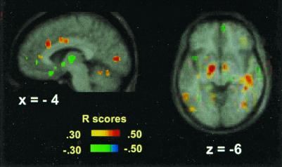
Figure 2.
Positive and negative correlations of breathlessness scores with rCBF during CO2 FM and CO2 MP scans displayed on the average MRI brain image. Significant correlations are evident in the lingual gyrus, anteriorly and rostrally in the cingulate gyrus (x = −4), and in the sublenticular area and right temporal gyrus (z = −6). The color coding of the correlation coefficients is shown.

