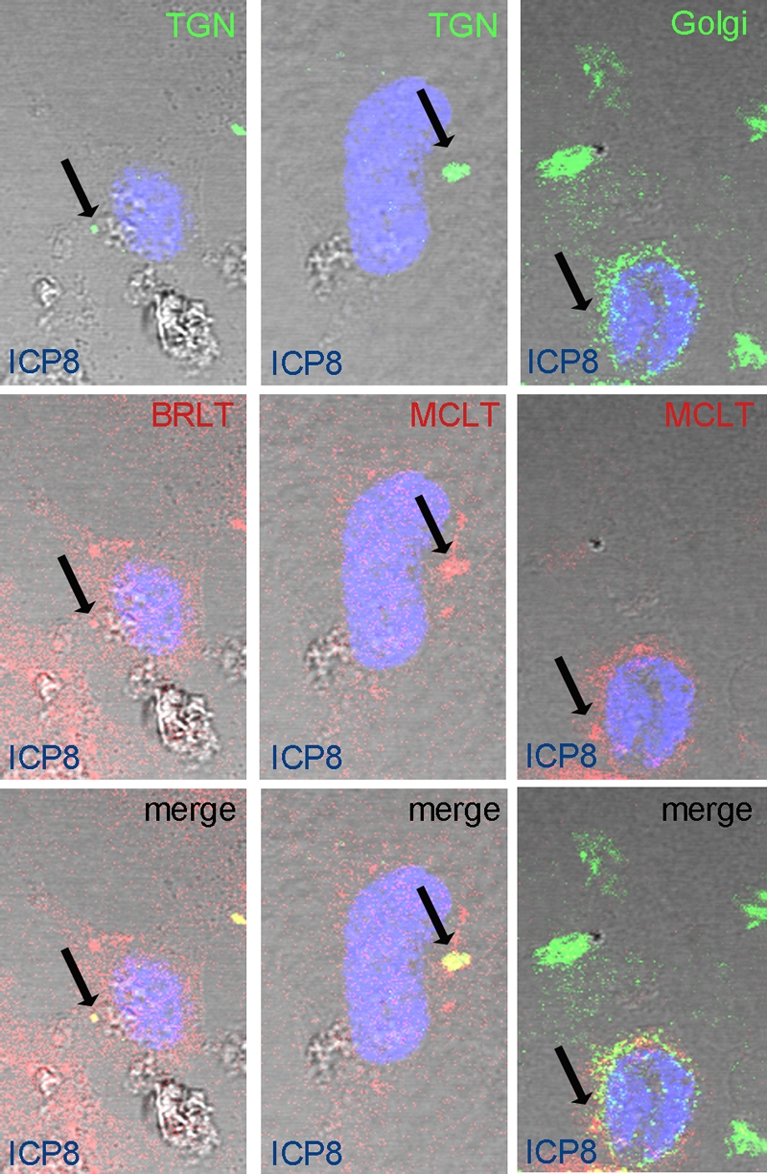
FIG. 4.

DN-MyoVa colocalizes with TGN but not Golgi markers in HSV-infected cells. HeLa cells were transfected with mRED-DN-myoVa BRLT (left column) or MCLT (center and right columns). At 23 h posttransfection the cells were infected with HSV-1(F) at an MOI of 5 PFU per cell. At 16 hpi the cells were washed with PBS, fixed in 3% PFA for 15 min, and permeabilized with 0.1% Triton X-100. Fixed cells were immunostained with mouse anti-TGN (top row, first and second columns) or mouse anti-Golgi markers (top row, right column). Rabbit anti-ICP8 was used to mark the infected cell nuclei. Red, DN-myoVa; green, TGN and Golgi; blue, ICP8. Arrows indicate sites of colocalization of the respective markers.
