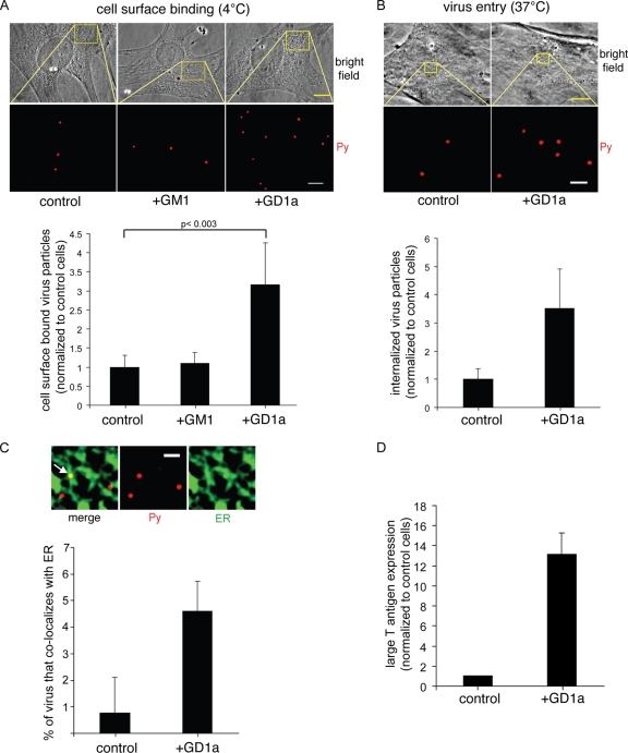
FIG. 1.
GD1a addition to a murine cell line lacking functional receptors stimulates Py binding, entry, ER transport, and infection. (A and B) Control cells, GM1-supplemented cells (shown in panel A alone), and GD1a-supplemented A1-1 cells were incubated with Py at 4°C for 1 h (A) or at 37°C for 4 h (B), washed to remove the unbound virus, and subjected to immunofluorescence with an antibody against VP1. Top panel, representative images. Scale bars, 10 μm for bright-field images and 2 μm (A) or 1 μm (B) for Py images. Bottom panel, quantification of Py binding to the plasma membrane from at least 3 cells. Data represent means ± standard deviations. A two-tailed t test was used. (C) Control and GD1a-supplemented A1-1 cells expressing CFP-HO2 were incubated with Py at 4°C for 40 min, washed to remove the unbound Py, and then incubated at 37°C for 4.5 h. Cells were subjected to immunofluorescence with an antibody against VP1. Top panels, representative images. Arrow, Py that colocalized with the ER. Scale bar, 1 μm. Bottom panel, quantification of the Py colocalizing with the ER from at least 3 cells. Data represent means ± standard deviations. (D) Control and GD1a-supplemented A1-1 cells were incubated with Py at 37°C for 48 h and subjected to immunofluorescence with an antibody against the large T antigen. Data represent means ± standard deviations of the results of at least 2 independent experiments. A total of 3 of 4,004 cells expressed T antigen in the control cells.

