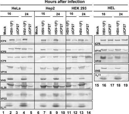
FIG. 5.
Viral protein accumulation in ΔICP27 virus-infected human cell lines. Confluent cell monolayers of HeLa (lanes 1 to 5), HEp-2 (lanes 6 to 10), HEK 293T (lanes 11 to 14), and HEL (lanes 15 to 19) cells were either mock infected (lanes 1, 6, and 15) or infected with 10 PFU per cell of HSV-1(F) (lanes 2, 4, 7, 9, 11, 13, 16, and 18) or the ΔICP27 mutant virus (lanes 3, 5, 8, 10, 12, 14, 17, and 19). The cells were harvested 16 h (lanes 2, 3, 7, 8, 11, 12, 16, and 17) or 24 h (lanes 4, 5, 9, 10 13, 14, 18, and 19) after virus exposure and processed as described in Materials and Methods. Equal amounts of proteins were electrophoretically separated on a 10% denaturing polyacrylamide gel, transferred to a nitrocellulose sheet, and reacted with the antibodies made against representative α (ICP4, ICP0), β (ICP8), or γ (VP16, VHS, VP22, UL38 and US11) proteins. Note that ICP4 forms multiple bands, reflecting posttranslational modifications of the protein.

