
FIG. 3.
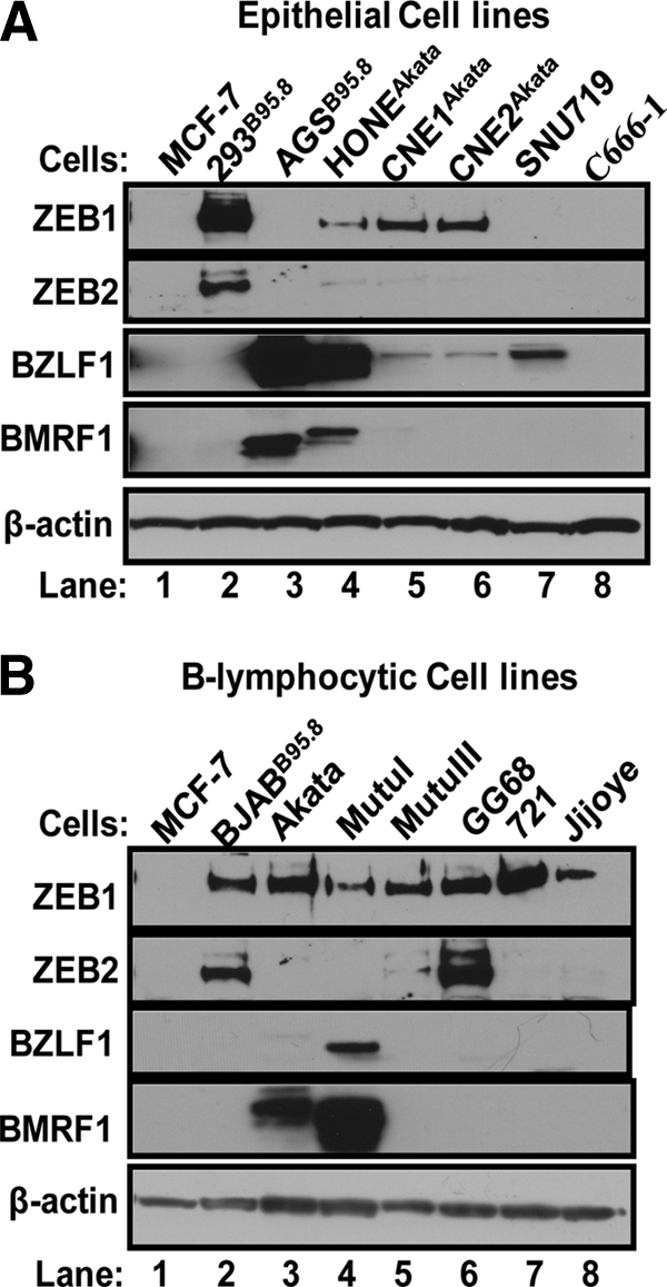
Immunoblots showing relative levels of ZEB1 and ZEB2 protein and the EBV IE BZLF1 and E BMRF1 proteins present in EBV-positive epithelial (A) and B-lymphocytic (B) cell lines. Thirty μg of whole-cell protein was loaded per lane and analyzed for relative levels of ZEB1, ZEB2, BZLF1, and BMRF1 protein as described in Materials and Methods. β-Actin served as a loading control.