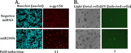
FIG. 7.
miR200b activates the full EBV lytic cycle in CNE1Akata cells. (A) Indirect immunofluorescence staining showing the addition of miR200b leads to induction of EBV late gene expression. CNE1Akata cells were transfected with 30 nM of the negative control or miR200b and incubated at 37°C for 72 h prior to processing for gp350 protein as described in Materials and Methods. Shown here are fields of cells observed for all cellular nuclei (Hoechst staining) versus the subset of them containing gp350 protein. (B) Raji cell assay showing increased production of infectious virus in CNE1Akata cells after the addition of miR200b. Media from cells transfected and incubated as described in panel A were concentrated and used to infect Raji cells as described in Materials and Methods. Shown here are fields of Raji cells observed with visible light (Light) for all cells versus UV light (UV) for the EBV-infected, GFP-positive ones.

