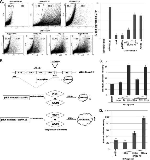
FIG. 4.
RNAi suppression activity of 7a in the mammalian cell line. (A) The left panel represents the FACS analysis of the mammalian cell line HEK293T transfected with 0.5 μg of RNAi-ReadypSIREN-RetroQ-ZsGreen-Retroviral vector (Clonetech) coexpressing GFP along with shRNA against GFP and viral ORFs (7a and FHV B2). All the ORFs were cloned downstream of the human cytomegalovirus (CMV) promoter in the vector pCDNA. The GFP expression of the cells was analyzed at 3 days posttransfection. The control GFP expression was monitored by replacing short hairpin GFP (shGFP) with short hairpin luciferase (shLuc), where 89%of the cells showed GFP fluorescence. The shGFP induced 79% reduction in GFP-expressing cells. The restoration of GFP silencing was monitored in the presence of increasing amounts (300 ng and 1 μg) of 7a as well as the positive control FHV B2. Each transfection experiment was carried out in three replicates. The right panel shows the bar graph representation of the FACS result, with the percentage of cells expressing GFP represented on the y axis and different transfection combinations represented on the x axis. (B) Schematics of the structural features of HIV-1 replicon DNA (pNL4-3.LucR-E-), showing the insertion of the firefly luciferase gene in the Nef-encoding region. Upon transfection into mammalian cells, the luciferase gene is expressed from a spliced transcript. The extent of luciferase is increased when the cells (293T or A549 cells) are cotransfected with the 7a construct, compared to the level for the control DNA (pCDNA). (C) Increase in HIV replicon activity obtained with the 7a protein. HEK293T cells were transfected with 100 ng of the HIV-1 clone pNL-LucR-E- and increasing concentrations of 7a (and the positive-control NS1 gene from influenza virus), along with 10 ng of the pTK-RL plasmid, using Lipofectamine reagent. At 48 h posttransfection, cell lysates were prepared in accordance with standard protocols and the luciferase values were measured using a dual luciferase kit from Promega. The relative luciferase amounts were monitored for the various transfection combinations and are represented in the bar diagram. All the values were normalized to those for Renilla luciferase. (D) Analysis of 7a activity in the SARS-CoV-permissive human lung epithelial cell line A549 by a replicon enhancement assay. One hundred nanograms of HIV-1 clone pNL-LucR-E was transfected with increasing concentrations of 7a by use of Lipofectamine reagent. At 48 h posttransfection, the cell lysate was prepared and luciferase values were measured using a dual luciferase kit from Promega as described for panel C.

