
FIGURE 6.
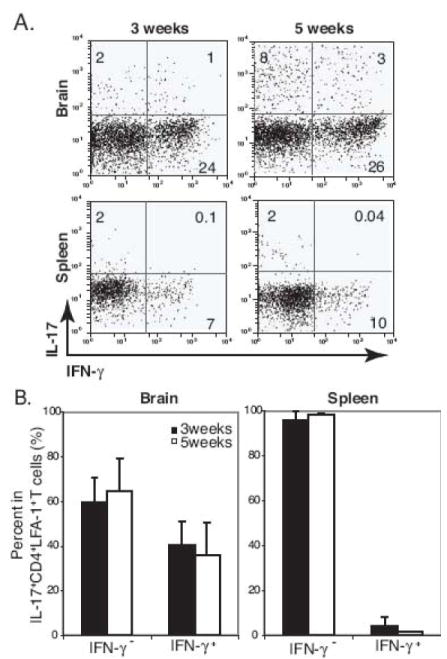
High frequency of IFN-γ+IL-17+ double-positive cells is detected in the brain during the course of i.c. BCG infection. A, Representative dot plots show IFN-γ+ and IL-17+-producing cells from a CD4+LFA-1high T cell gate from the brain and spleen at 3 and 5 weeks post i.c. BCG infection. Numbers represent the frequency of each quadrant among CD4+LFA-1high T cell cells. B, Summary graphs show the proportion of IFN-γ−IL-17+ and IFN-γ+IL-17+ cells in CD4+LFA-1highIL-17+ T cells from the brain and spleen at 3 and 5 weeks post i.c. BCG infection. Three to six mice were used per group.