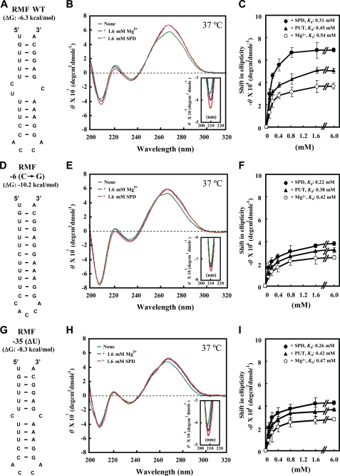
FIGURE 5.
CD spectra of RMF WT RNA, RMF −6(C→G) RNA, and RMF −35(ΔU) RNA. A, D, and G, structure of RMF WT RNA, RMF −6(C→G) RNA, and RMF −35(ΔU) RNA. B, E, and H, CD spectra were recorded as described under “Experimental Procedures.” Green line, no addition; blue line, 1.6 mm Mg2+; red line, 1.6 mm spermidine. C, F, and I. Concentration-dependent shifts induced by Mg2+ (○), putrescine (▴), and spermidine (●) at 37 °C in magnitude at 208 nm are shown. Values are means ± S.E. of triplicate determinations. The Kd values of spermidine, putrescine, and Mg2+ for RMF WT RNA, RMF −6(C→G) RNA, and RMF −35(ΔU) RNA at 37 °C were determined according to the double-reciprocal equation plot.

