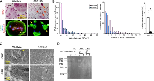
FIGURE 4.
Essential roles of CCR1 in multinucleation and bone-resorbing activity. Pre-osteoclastic cells were cultured from the bone marrow of wild-type and Ccr1−/− mice. Osteoclasts were induced from the pre-osteoclastic cells by M-CSF and RANKL treatment. In A, the formation of multinuclear osteoclasts by wild-type and Ccr1−/− precursors was visualized by TRAP chromogenic staining (magnification ×400, upper panels). Immunohistochemical staining was carried out using an anti-cathepsin K antibody conjugated with Alexa594 (red). F-actin and nuclei were counterstained by phalloidin-AlexaFluor 488 (green) and Hoechst 33258 (blue), respectively (magnification ×640, bottom panels). The yellow arrow indicates multinuclear giant cells with an impaired actin ring rearrangement, and the red arrows indicate TRAP accumulation. In B, histograms of the area distribution of multinuclear osteoclasts delimited with phalloidin, and of the number of multinuclear osteoclasts in A. Area comprises TRAP-positive multinuclear (>3 nuclei) giant cells shown in A (mean ± S.E., n = 3). In C, pit formation by wild-type and Ccr1−/− osteoclasts on bone slice observed by scanning electron microscopy (magnification: ×1000 (top) and ×6000 (bottom), respectively). In D, collagen digestion activity by wild-type and Ccr1−/− osteoclasts was measured by collagen-based zymography. Lanes M, 1, 2–3, and 4–5 indicate the molecular markers, bone marrow-derived macrophage lysates (10 μg of protein/lane), wild-type osteoclast lysates (1 and 10 μg of protein/lane), and Ccr1−/− osteoclasts lysates (1 and 10 μg of protein/each lane), respectively.

