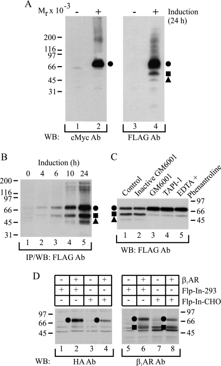
FIGURE 1.
hβ1AR is susceptible to N-terminal cleavage. A, receptor species expressed after long term induction. HEK293i cells stably transfected with the N-terminally Myc-tagged and C-terminally FLAG-tagged hβ1AR were induced with 0.5 μg/ml tetracycline for 24 h (lanes 2 and 4) or not (lanes 1 and 3). Isolated membranes were solubilized in SDS-sample buffer and subjected to Western blotting, using either anti-c-Myc (lanes 1 and 2) or anti-FLAG M2 (lanes 3 and 4) antibodies (Ab). B, time-dependent induction of receptor expression. Stably transfected HEK293i cells were induced for 0, 4, 6, 10, or 24 h, and the receptors were solubilized in DDM buffer, subjected to anti-FLAG M2 antibody immunoprecipitation, and analyzed by Western blotting with the same antibody. C, in vitro proteolysis. Stably transfected HEK293i cells were induced for 24 h, and cellular membranes were isolated using 25 mm Tris-HCl, pH 7.4, containing 2 μg/ml aprotinin, 0.5 mm phenylmethylsulfonyl fluoride, 5 μg/ml leupeptin, 5 μg/ml trypsin inhibitor, and 10 μg/ml benzamidine (lane 1). The buffer was supplemented with 10 μm inactive GM6001 (lane 2), 10 μm GM6001 (lane 3), 10 μm TAPI-1 (lane 4), or 2 mm EDTA and 2 mm 1,10-phenanthroline (lane 5). The DDM-solubilized receptors were subjected to anti-FLAG M2 antibody immunoprecipitation in the presence of the respective reagents, and the purified samples were analyzed by Western blotting with the same antibody. D, receptor expression in transiently transfected cells. The N-terminally HA-tagged hβ1AR was transiently transfected into Flp-In-293 (lanes 2 and 6) or Flp-In-CHO (lanes 4 and 8) cells. Control cells (lanes 1, 3, 5, and 7) were transfected with vector DNA. The receptors were solubilized in DDM buffer and subjected to Western blotting, using either the anti-HA antibody (lanes 1–4) or the anti-hβ1AR antibody (lanes 5–8), which is directed against the C terminus of the receptor. The full-length receptor is indicated with a closed circle and the proteolytic fragments with a closed square and a triangle. Molecular weight markers are indicated. IP, immunoprecipitation; WB, Western blotting.

