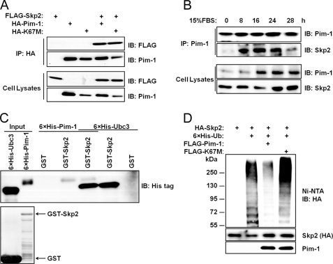
FIGURE 2.
Pim-1 binds to Skp2. A, HEK293T cells were transfected with the indicated plasmids, protein immunoprecipitated (IP) with HA antibody and immunoblotted with FLAG or Pim-1 antibody. Lysates of cells used for this assay are probed with identical antibodies. B, HEK293T cells were serum-starved (0.2%) for 48 h prior to the addition of 15% FBS at 0 time. Cells were then harvested at the indicated time points, and coimmunprecipitation (co-IP) was performed. C, GST-Skp2 proteins were incubated overnight with His-tagged Pim-1 or Ubc3 proteins purified from E. coli at 4 °C, washed with PBS, and subjected to immunoblot analysis (upper panel). GST and GST-Skp2 were stained with Coomassie Brilliant Blue (lower panel). D, HEK293T cells were transfected with the indicated plasmids, treated with 10 μm MG132 for 6 h, and ubiquitination as measured by binding to nickel-nitrilotriacetic acid (Ni-NTA) beads (see “Experimental Procedures”) followed by an immunoblot with anti-HA antibody. Immunoblot (IB) analysis was performed on total cell lysates from these HEK293T cells (two lower panels).

