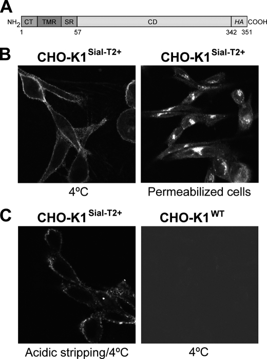
FIGURE 1.
Immunofluorescence detection of Sial-T2 in CHO-K1 cells. A, schematic representation of the Sial-T2-HA construct used to generate clone 2 (CHO-K1Sial-T2+) is shown. CT, cytoplasmic tail; TMR, transmembrane region; SR, stem region; CD, catalytic domain; and HA, nanopeptide epitope of the viral hemagglutinin. B, Sial-T2-HA-expressing CHO-K1 cells (CHO-K1Sial-T2+) were immunostained with an antibody to HA at 4 °C for 45 min and then fixed and incubated with a secondary antibody conjugated to Alexa Fluor 488 (4 °C, left panel). Alternatively, CHO-K1Sial-T2+ cells were fixed and permeabilized before immunostaining with antibody to HA and secondary antibody conjugated to Alexa Fluor 488 (Permeabilized cells, right panel). C, CHO-K1Sial-T2+ cells were incubated with acetate buffer at 4 °C for 1 min before immunostaining with antibody to HA and secondary antibody conjugated to Alexa Fluor 488 (Acidic stripping/4 °C, left panel). Wild-type CHO-K1 cells (CHO-K1WT) were incubated at 4 °C with antibody to HA at 4 °C for 60 min and then fixed and incubated with secondary antibody conjugated to Alexa Fluor 488 (4 °C, right panel). The image contrast in CHO-K1WT cells was reduced to show the presence of cells. Single confocal sections were taken every 0.7 μm parallel to the coverslip.

