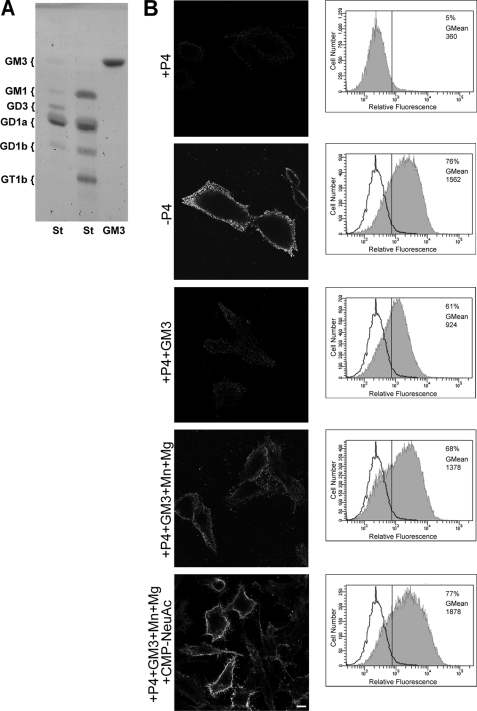
FIGURE 3.
Ecto-Sial-T2 sialylates exogenously incorporated GM3 in CHO-K1Sial-T2+ cells. A, chromatographic analysis of GM3 ganglioside used in the experiments in B is shown. GM3 and standard (St) glycolipids were co-chromatographed on HPTLC and revealed by orcinol staining. The positions of glycolipid standards are indicated on the left. B, CHO-K1Sial-T2+ cells were grown with P4 (+P4; first and third to fifth rows) or without P4 (−P4, second row) for 4 days. Then, cells were treated with 100 μm GM3, washed, and incubated at 37 °C for 2 h in a medium containing only DMEM (+P4+GM3, third row) or in a medium containing Mn2+ and Mg2+ (+P4+GM3+Mn+Mg, fourth row) or CMP-NeuAc, Mn2+ and Mg2+ (+P4+GM3+Mn+Mg+CMP-NeuAc, fifth row). P4 inhibitor remained present throughout the experiments. Left panels, cells were washed, immunostained with antibody to GD3 (R24) at 4 °C for 60 min, and then fixed and incubated with secondary antibody conjugated to Alexa Fluor 488. Single confocal sections were taken every 0.7 μm parallel to the coverslip. Right panels, cells were trypsinized, incubated at 4 °C with R24 antibody for 30 min, and then fixed and exposed to the secondary antibody for 30 min at 4 °C. Labeled cells were washed, resuspended in 200 μl of PBS, and fluorescence-quantified using flow cytometric analysis. The vertical line in each histogram marks the upper limit of control (+P4) to assess frequencies (%) of positive cells. The geometric mean fluorescence intensity (GMean) is also indicated. Scale bar, 10 μm.

