FIGURE 4.
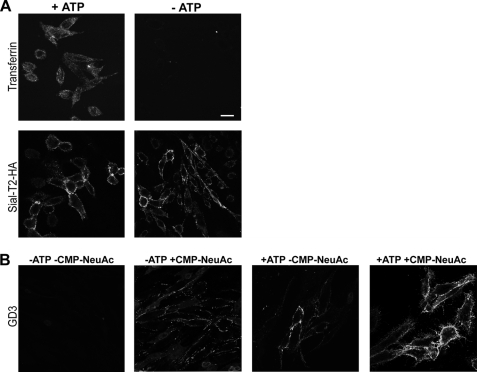
Inhibition of vesicular transport reduces GD3 synthesis at the cell surface of CHO-K1Sial-T2+ cells. A, CHO-K1Sial-T2+ cells were incubated for 1 h in PBS containing 50 mm 2-deoxyglucose and 5 mm NaN3 (−ATP) or complete DMEM (+ATP). Cells were labeled with Alexa Fluor 647-transferrin and then fixed and visualized by confocal microscopy (upper panels). Alternatively, cells were immunostained with antibody to HA at 4 °C for 1 h (Sial-T2-HA) and then fixed and incubated with secondary antibody conjugated to Alexa Fluor 488. Single confocal sections of 0.7 μm were taken parallel to the coverslip. B, CHO-K1Sial-T2+ cells were treated with P4 for 4 days and incubated for 3 h with 100 μm GM3. Then, cells were incubated for 1 h in PBS containing 5 mm NaN3 and 50 mm 2-deoxy-d-glucose (−ATP), or complete DMEM (+ATP). Finally, cells were incubated for 2 h at 37 °C in a medium containing P4, Mn2+, and Mg2+, both in the presence (+CMP-NeuAc, second and fourth panels) or in the absence of CMP-NeuAc (−CMP-NeuAc, first and third panels). Then, cells were washed and immunostained with R24 antibody to GD3. Single confocal sections were taken every 0.7 μm parallel to the coverslip. Scale bar, 10 μm.

