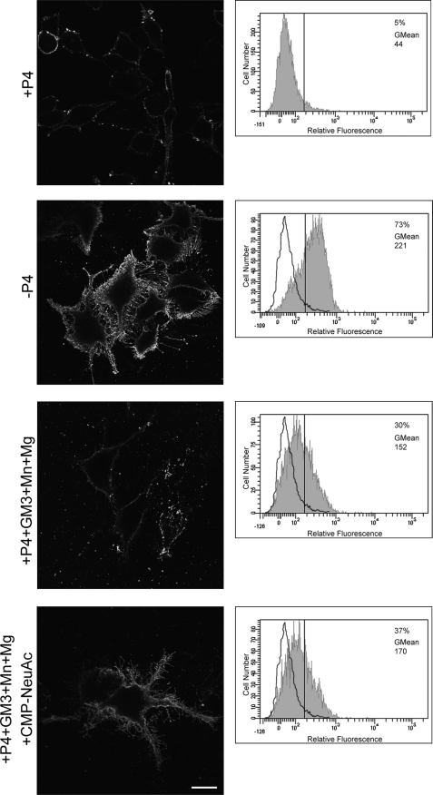
FIGURE 5.
GD3 synthesis at the cell surface of SK-Mel-28 human cells endogenously expressing Sial-T2. SK-Mel-28 cells were grown with P4 (+P4; first, third, and fourth rows) or without P4 (−P4, second row) for 4 days. Then, cells were treated with 100 μm GM3, washed, and incubated at 37 °C for 2 h in a medium containing Mn2+ and Mg2+ (+P4+GM3+Mn+Mg, third row), or CMP-NeuAc, Mn2+ and Mg2+ (+P4+GM3+Mn+Mg+CMP−NeuAc, fourth row). P4 inhibitor remained present throughout the experiments. Left panels, cells were washed, immunostained with antibody to GD3 (R24) at 4 °C for 1 h and then fixed and incubated with secondary antibody conjugated to Alexa Fluor 488. Single confocal sections were taken every 0.7 μm parallel to the coverslip. Right panels, cells were trypsinized, incubated at 4 °C with R24 antibody for 30 min, and then fixed and exposed to the secondary antibody for 30 min at 4 °C. Labeled cells were washed and resuspended in 200 μl of PBS, and fluorescence was quantified using flow cytometric analysis. The vertical line in each histogram marks the upper limit of control (+P4) to assess frequencies (%) of positive cells. The geometric mean fluorescence intensity (GMean) is also indicated. Scale bar, 10 μm.

