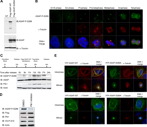
FIGURE 4.
Phosphorylation of ASAP at Ser-289 by Plk1 in vivo occurs at centrosomes during mitosis. A, U-2 OS cell lysates transfected with FLAG-ASAP-WT or FLAG-ASAP-S289A were immunoblotted (IB) with the anti-ASAP-P-S289 (left) and anti-ASAP (right) antibodies. NT, nontransfected cells. α-Tubulin is shown as a loading control. B, asynchronous U-2 OS cells were grown on glass coverslips, fixed in F/PHEM/methanol, probed with the anti-ASAP-Ser(P)-289 (green) and anti-γ-tubulin (red) antibodies, and stained with Hoechst 33258 (blue); the different stages of the cell cycle are shown from G2 to telophase (scale bar, 10 μm). C, U-2 OS cells were synchronized by double thymidine block and transfected with FLAG-ASAP during the first release, as indicated on the schematic. At the indicated time points after the second release, cells lysates were analyzed by immunoblotting using the indicated antibodies. α-Actin was used as a loading control. D, U-2 OS cells were transfected with FLAG-ASAP-WT, synchronized by thymidine block, and released in the presence of either dimethyl sulfoxide (DMSO) (control) or 1 μm TAL for 13 h. Cell extracts were immunoblotted with the anti-ASAP-Ser(P)-289, anti-FLAG, anti-Plk1, and anti-H3-Ser(P)-10 antibodies. α-Actin is shown as a loading control. E, U-2 OS cells were grown on glass coverslips, transfected with YFP-ASAP-WT (left panels) or YFP-ASAP-S289A mutant (right panels), fixed in PAF/MTSB (upper panels) or in F/PHEM/methanol (lower panels), and probed with anti-ASAP (green) and anti-α-tubulin (red) (top), anti-γ-tubulin (red) (bottom) and stained with Hoechst 33258 (blue) (scale bar, 10 μm).

