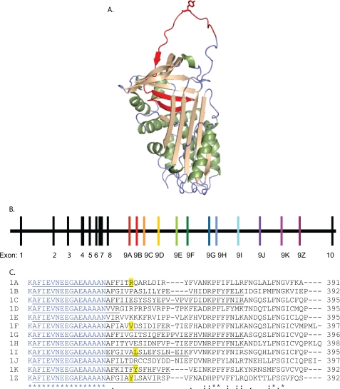
FIGURE 1.
Mutually exclusive splicing of the serpin-1 gene to include different versions of exon 9 produces serpin isoforms with different reactive center loop sequences. A, protein structure of serpin-1K (PDB code 1SEK). The alternatively spliced region is shown in red, and the P1 residue, Tyr359, is shown in the “stick” format. B, structure of the M. sexta serpin-1 gene. The first 8 exons encode the constant region of the protein, which is followed by the carboxyl-terminal variable region, encoded by an exon 9. Each exon 9 includes a stop codon to end the open reading frame. Exon 10 includes the 3′ untranslated region. C, an alignment of the carboxyl termini of the 12 serpin-1 isoforms starting with Lys336. The region encoded by the 3′ end of exon 8 is in blue; exon 9 starts after Asn352. Known P1 residues are highlighted in yellow. The first isoform-specific tryptic peptide for each isoform is underlined.

