Figure 4.
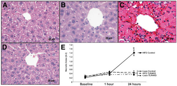
TLR4 deficiency reduces hepatocellular necrosis following ischemia/reperfusion. Prior to surgery and 1 and 24 hours after reperfusion, H&E slides were made from tissue harvested from the left hepatic lobe of each mouse. At 24 hours following reperfusion, no appreciable necrosis was observed in (A) control ND or (B) TLR4KO ND groups, as illustrated by the representative photomicrographs. (C) However, necrosis was marked in the control HFD group, in which large necrotic foci were present around central veins, whereas (D) TLR4 HFD animals showed little appreciable necrosis. (E) Grading of centrilobular necrosis for each animal was conducted on a 0 to 3 scale, scored per high-powered field, and represented graphically. At 24 hours, necrosis was significantly greater in the control HFD livers compared to the TLR4KO HFD livers, whereas it was greater for both groups in comparison with their ND controls. Values are expressed as the mean ± the standard error of the mean (*P < 0.05 versus HFD TLR4KO, n = 5–8/group). Abbreviations: HFD, high-fat diet; ND, normal diet; TLR4, toll-like receptor 4; TLR4KO, toll-like receptor 4–deficient.

