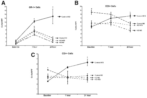
Figure 5.
TLR4 deficiency decreases cellular infiltration following ischemia/reperfusion. (A) Sections were stained for GR-1, a granulocyte marker for neutrophils. All groups had increased infiltration at baseline (§P < 0.05 versus baseline). Additionally, there was a significant increase in the neutrophil infiltration in control HFD animals versus TLR4KO HFD animals at 1 hour of reperfusion(*P < 0.05, n = 5–8/group). As the ND groups and the TLR4KO HFD groups returned to baseline levels at 24 hours, the control HFD animals retained a significantly elevated level of neutrophil infiltration (*P < 0.05, n = 5–8/group). (B) At baseline, there were fewer CD3+ cells in the control HFD animals versus the control ND animals (ψP < 0.05). This trend continued when the control ND animals were compared with the TLR4KO ND animals at 1 hour (ψP < 0.05). At 24 hours, the control HFD group was significantly increased over both knockout groups (ζP < 0.05). (C) There was a similar trend at baseline in the CD4+ cells, in that the control HFD animals had significantly fewer cells than the control ND animals (*P < 0.05). There were no differences at 1 hour of reperfusion, but by 24 hours, there were more infiltrating cells in the control HFD animals than in the TLR4KO HFD animals (*P < 0.05). Values are represented as the mean number of cells per HPF ± the standard error of the mean. Abbreviations: HFD, high-fat diet; HPF, high-powered field; KO, knockout; ND, normal diet; TLR4, toll-like receptor 4; TLR4KO, toll-like receptor 4–deficient.

