Figure 3.
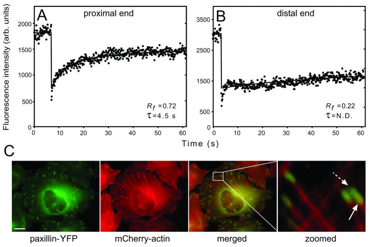
Paxillin and vinculin display different dynamics at the FA proximal and distal ends. FRAP experiments were carried out, focusing the beam on the two FA ends. (A) A typical FRAP curve of paxillin-YFP at the proximal (heel) FA end (60 s timescale). (B) A typical curve at the distal (toe) FA end (60 s timescale). Fast recovery by diffusion exists, but the ensuing exchange is very slow. (C) Paxillin co-localizes with actin at the FA proximal end. HeLa-JW cells co-expressing paxillin-YFP and mCherry-actin were visualized by fluorescence microscopy. Co-localization was visible at the proximal edge (solid arrow), but not at the distal edge (dashed arrow). Scale bar: 10 μm. [Adapted from Wolfenson et al. 2009]

