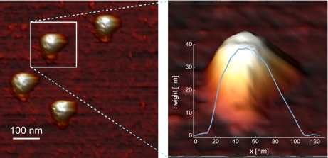
Fig. 3.
Sample AFM image of noroviruses in liquid. On the left is a top view image showing several viral particles. In this view, structural details of the particles (cuplike depressions) can already be recognized. On the right is a zoomed-in side view image on one of the particles shown in a three-dimensional representation. The cross-sectional profile shows the apparent overestimation of the lateral dimensions of the sphere-like particle, which results from the tip convolution effect inherent to AFM imaging (80, 83). However, the diameter can be accurately determined by measuring the height. The z-profile indicates a maximum height of about 38 nm, which is in close agreement to the diameter determined by electron microscopy and x-ray crystallography (84, 85). The images were recorded using jumping mode AFM (see Ref. 86 for full experimental details with the sole difference that these images were taken in sodium phosphate buffer).

