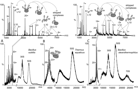
Fig. 2.
Mass spectra of 70 S ribosomes from B. subtilis, T. aquaticus, and B. stearothermophilus ribosomes and tandem MS of two pentameric stalk complexes. Mass spectra and tandem MS spectra of 70 S ribosomes from B. subtilis (A), T. aquaticus (B), and B. stearothermophilus (C) ribosomes in 1 m ammonium acetate, pH 7.5 are shown. Conditions were optimized for observation of the stalk complex. Individual charge states of two stalk complexes from B. subtilis and B. stearothermophilus were then isolated in the quadrupole and accelerated in the collision cell to analyze their components (MS/MS or tandem MS) (upper panels). At m/z values lower than the isolated peak individual, highly charged proteins are observed that have been released from the complex. At m/z values higher than the isolated charge state peaks are assigned to complexes “stripped” of these released proteins. The schematics at the top of the figure illustrate this process: L10 is represented in dark gray, and L12 composed of two domains connected via a flexible linker is represented in light gray. MS/MS spectra above 3000 m/z have been magnified by a factor of 5 in both cases.

